Autore: Mihai Nan
În această problemă se urmărește dezvoltarea unui model de clasificare a imaginilor capabil să distingă între semnale ECG normale și ECG anormale, pornind de la un set de date cu informații medicale reale.
Datele furnizate în cadrul acestei probleme sunt reprezentate sub forma unor imagini ECG, fiecare imagine corespunzând unei înregistrări complete a activității electrice a inimii pentru un pacient. Imaginile pot fi utilizate direct ca date de intrare pentru modele de clasificare a imaginilor.
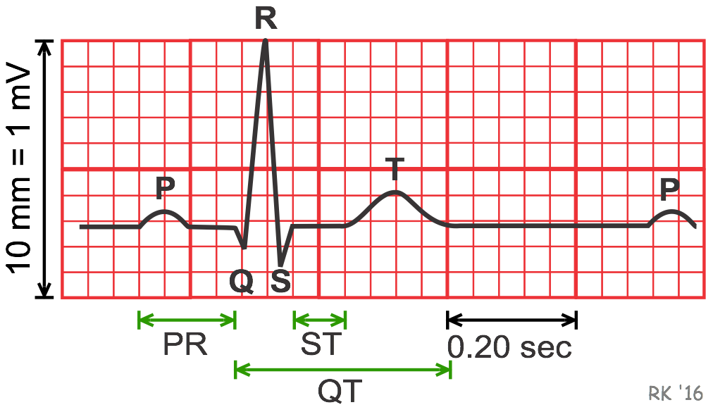
ECG (Electrocardiograma) este o investigație medicală neinvazivă care înregistrează activitatea electrică a inimii în timp.
Fiecare bătaie a inimii este controlată de impulsuri electrice, iar ECG-ul măsoară aceste semnale folosind electrozi plasați pe suprafața corpului.
ECG-ul este utilizat pe scară largă pentru:
Anomaliile din semnalul ECG pot indica afecțiuni cardiace grave, motiv pentru care automatizarea procesului de analiză este extrem de importantă în practica medicală.
Setul de date utilizat conține înregistrări ECG preprocesate, cu următoarele caracteristici:
0 – ECG normal1 – ECG anormalDatele sunt organizate în fișiere CSV:
train.csv – imagini și etichete pentru antrenaretest.csv – imagini fără etichete (pentru inferență)Scopul este de a construi un model de clasificare a imaginilor care, pe baza unei imagini ECG, să prezică dacă semnalul cardiac este normal sau anormal.
Modelul trebuie să:
La final, vei genera un fișier submission.csv cu forma:
image_path,labelimages/ecg_000001.png,0images/ecg_000002.png,1Metrica folosită pentru evaluarea modelului este acuratețea:
accuracy = (număr_predicții_corecte / număr_total_predicții)